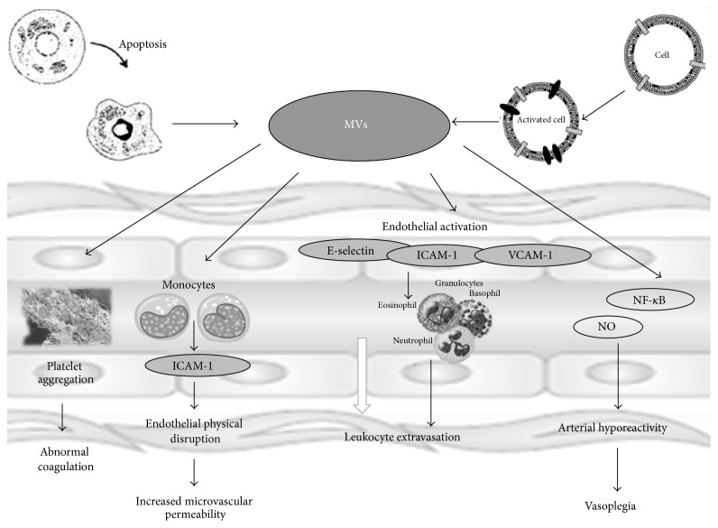
Figure 3.
A schematic representing microvesicles (MVs) functions. MVs are released in the extracellular environment through a membrane reorganization and blebbing process following cell activation or apoptosis. They contribute to the spread of inflammatory and prothrombotic vascular status and they may affect smooth muscle tissue through adhesion molecules (E-selectin, ICAM, and VCAM) activation of nuclear factor κB and the expression of inducible nitric oxide synthase and cyclooxygenase-2, with an increase in nitric oxide (NO) and vasodilator prostanoids, leading to arterial hyporeactivity.

