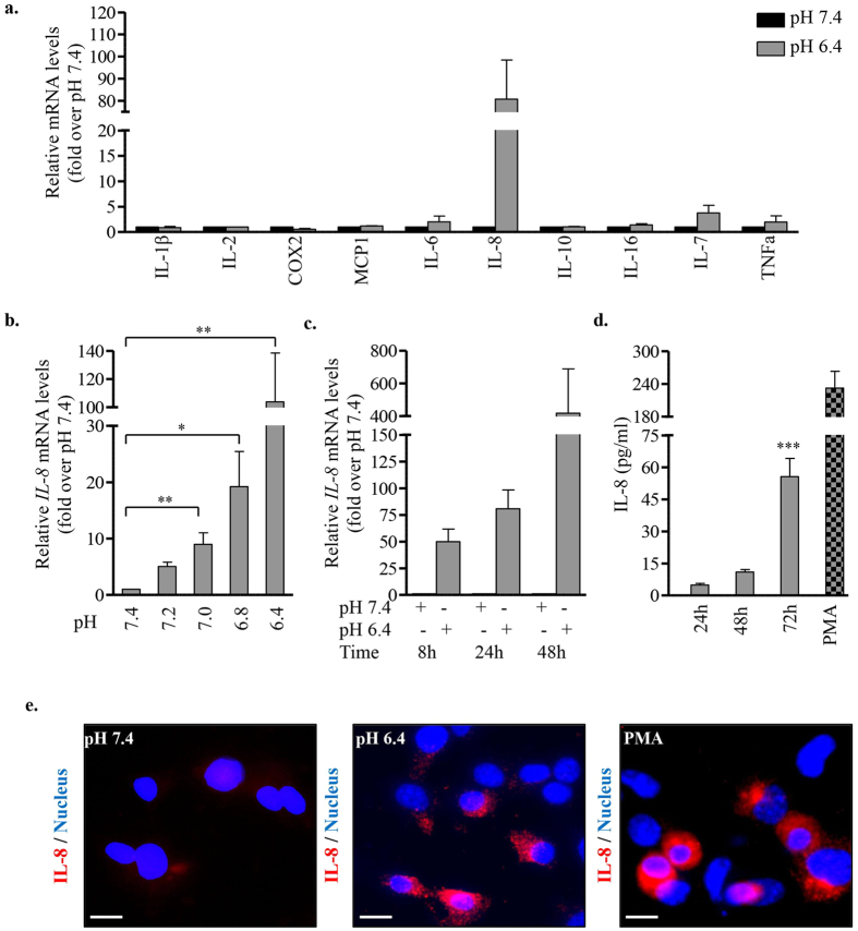
Figure 4. Acidic extracellular pH induces the expression and secretion of IL-8 by EndoC-βH2 cells.
(a) EndoC-βH2 cells were cultured at pH 7.4 or 6.4 for 24 h and screened by RT-qPCR for the expression of selected pro/anti-inflammatory cytokines. (b) IL-8 expression was determined by RT-qPCR in EndoC-βH2 cells cultured at different pH for 24 h (one-way ANOVA, post test for linear trend (p = 0.0008)). (c) IL-8 expression was determined by RT-qPCR in EndoC-βH2 cells cultured at pH 7.4 or 6.4 for the indicated time points (one-way ANOVA, post test for linear trend (p = 0.067)). (d) EndoC-βH2 cells were cultured at pH 7.4 or 6.4 for 24 h/48 h/72 h or with PMA (100 ng/ml for 12 h) (one-way ANOVA). Secreted IL-8 was quantified by ELISA. (e) Immunostaining for IL-8 in EndoC-βH2 cells cultured at pH 7.4 or 6.4 for 72 h or with PMA (100 ng/ml) for 12 h. Scale bar 10 μm. Data are mean ± SEM of 3–5 experiments. *p < 0.05; **p < 0.01; ***p < 0.001.

