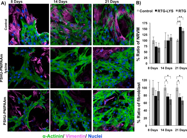
Figure 4.
Fluorescence staining of NRVM and fibroblast growing in different substrates. sarcomeric α-actinin (green), vimentin (pink), and DAPI (blue). (A) Top-row panels: NRVM cultured on 2D tissue culture plate coated with gelatin (control) after 8, 14, and 21 days. Middle-row panels: NRVM cultured in 3D PSHU-PNIPAAm-lysine after 8, 14, and 21 days. Bottom-row panels: NRMV cultured in 3D PSHU-PNIPAAm after 8, 14, and 21 days. Compared to controls, the 3D matrices stimulate NRVM to spread and form heart-like functional syncytia. (B) Quantification histogram of the ratio of NRVM and fibroblast growing in gelatin-control and RTG biopolymers. Values were normalized to controls. Significant differences on the % ratio of NRVM can be observed after 21 days between gelatin control group and RTG biopolymers. ** indicates p value < 0.01 (Student’s test) (PNIPAAM-lysine p value: 0.0005; n = 5; PSHU–PNIPAAM p value: 0.002; n = 5). Significant differences on the % ratio of fibroblast was also observed after 14 and 21 days between gelatin control group and RTG biopolymers. * indicates p value < 0.05 (Student’s test; 14 days: PSHU-PNIPAAM-lysine p value: 0.013; n = 5; PSHU-PNIPAAM p value: 0.02; n = 5. 21 days: PSHU-PNIPAAM-lysine p value: 0.023; n = 5; PSHU–PNIPAAM p value: 0.01; n = 5). Data are presented as mean ± SD (n = 5).

