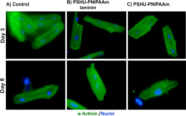
Figure 8.
Fluorescence sarcomeric α-actinin (green) and DAPI (blue) staining of ARVM in different conditions: (A) 2D tissue culture plate coated with laminin (control), (B) 3D PSHU-PNIPAAm-laminin, and (C) 3D PSHU-PNIPAAm. Top-row panels ARVM after 3 days of culture. Bottom-row panels ARVM after 8 days of culture. Compared to control groups we found that the 3D PSHU-PNIPAAm-laminin matrix allows a long-term ARVM survival with a well-defined cardiac phenotype represented by a sarcomere striation.

