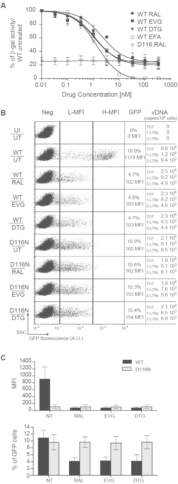
Figure 1. Expression of uDNA depending on DNA topology and integrase functionality.

(A) Level of integrated HIV-1 LTR transactivation depending on Tat expression from integrated or unintegrated viral DNA. Hela-P4 cells were infected with HIV NL4-3 virus harboring a wild-type (WT) integrase or a catalytically defective integrase (“D116N”) (20 ng of p24gag antigen on 106 cells; ≈m.o.i. 0.2) in the presence of various concentrations of INSITs (raltegravir, RAL; dolutegravir, DTG; elvitegravir, EVG) or reverse transcriptase inhibitor (efavirenz, EFA). β-galactosidase expression was quantified 72 h post-infection by the CPRG method (see Methods for details). The results are presented as the percentage of reporter gene expression relative to the activity measured after infection by NL4.3 WT virus in untreated condition (without drug). Results were obtained from 3 independent experiments (mean ± s.e.m). (B,C) Cytometric analysis of viral transgene expression depending on viral integrase activity. MT4 T-cells were infected with either HIV-1 env–gfp+ harboring a wild-type (WT) integrase or HIV-1 with a catalytically defective integrase (“D116N”), each pseudotyped with VSV-G protein (4 ng of p24gag antigen on 106 cells; ≈m.o.i. 0.1), in the absence (UT) or presence of 200 nM RAL, DTG or EVG. Three days later, cytofluorometry was employed to determine the percentage and mean fluorescence intensity (MFI) of GFP expression. Dot plots show GFP fluorescence (expressed in arbitrary units, A.U.) depending on the Side-Scatter light (SSC). Quantitative-PCRs directed against viral DNAs (total, 1-LTRc and 2-LTRc) were analyzed 72 h post-infection. One representative experiment (panel (B)) and the results of 4 independent experiments (mean ± confidence intervals for a p value < 0.05 - panel (C)) are presented. Other abbreviations: H-MFI: high MFI, L-MFI: low MFI; Neg: negative cells; UI: uninfected condition; UT: untreated cells.
