Figure 9.
Manganese Supplementation Partially Restores Photosynthesis in the cgld1 Mutant of Chlamydomonas.
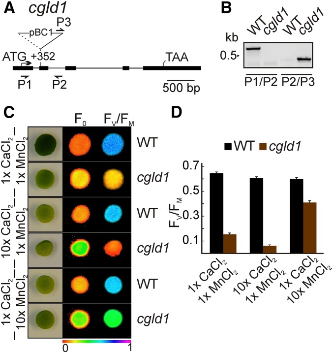
(A) Tagging of the CGLD1 locus (mRNA model XM_001701863). Exons are shown as black boxes and introns as black lines. Start and stop codons are indicated. The cgld1 allele was identified by Dent et al. (2015) (CAL029_02_05). The location of the insertion is indicated with respect to the start codon. The insertion is not drawn to scale. Binding sites of primers used for genotyping are indicated by arrows.
(B) The position of the insertion of the paromomycin resistance cassette was verified by PCR using wild-type (P1/P2) and mutant-specific (P2/P3) primer pairs.
(C) Test of PSII function in the presence of increased CaCl2 and MnCl2 concentrations. Cells were grown photoheterotrophically after spotting (105 cells per spot) onto TAP plates containing 0.34 mM (1×) or 3.4 mM (10×) CaCl2 and 25 µM (1×) or 250 µM (10×) MnCl2 in the indicated combinations, and the minimal chlorophyll a fluorescence (F0) (middle panel) and maximum quantum yield of PSII (FV/FM) (right panel) were recorded. The colored scale at the bottom indicates the signal intensities.
(D) FV/FM values are indicated for the wild type and cgld1 grown on the indicated TAP medium in (C). Bars represent values from five biological replicates (±sd).

