Figure 9.
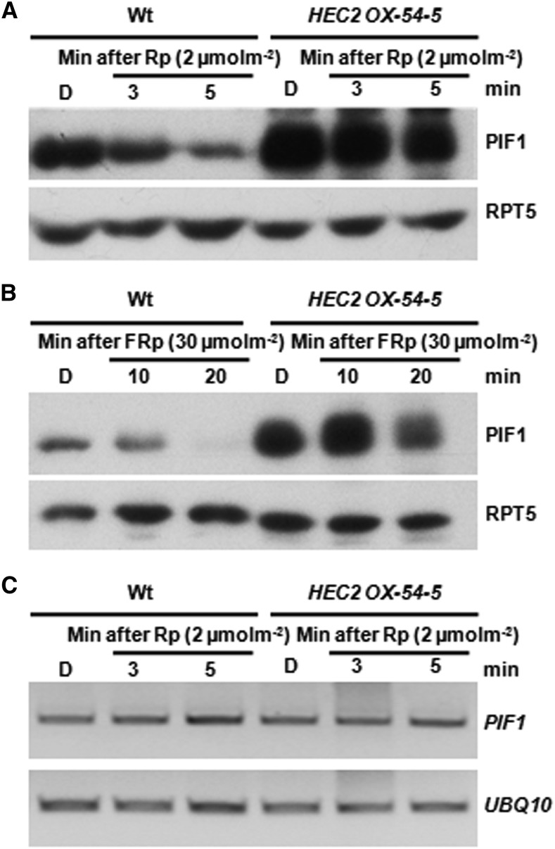
HEC2 Reduces the Light-Induced Degradation of PIF1.
(A) and (B) Immunoblots showing PIF1 level in the HEC2 OX line and wild-type Col-0. Four-day-old dark-grown seedlings were either kept in darkness or exposed to R (A) or FR (B) pulse and incubated in the dark for the time indicated. Total protein was extracted, separated in 8% SAD-PAGE gel, and transferred to PVDF membranes. Native PIF1 antibody was used to detect PIF1 protein level and anti-RPT5 antibody was used as control. Amount of light pulse is shown on the figure.
(C) The expression of PIF1 was not altered upon light treatment. RNAs were extracted from samples under the same treatment as in (A) and reverse transcribed. RT-PCR was performed to detect PIF1 expression level. UBQ10 was used as control.

