
Figure 2.
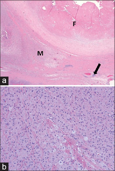
70-year-old man with increasing left hip pain diagnosed with high wear of a metal-on-metal total hip arthroplasty requiring left hip arthroplasty revision surgery. (a) Light microscopy (H and E, ×2.5) shows replacement of the synovial surface by organizing fibrin (F), prominent infiltrates of macrophages (M) with variable amounts of metallic wear debris and scattered perivascular lymphocyte aggregates (arrow). (b) Light microscopy (H and E, ×20) high power view of the perivascular lymphocyte aggregate indicated by the arrow in (a) shows prominent infiltrate of macrophages containing wear particles