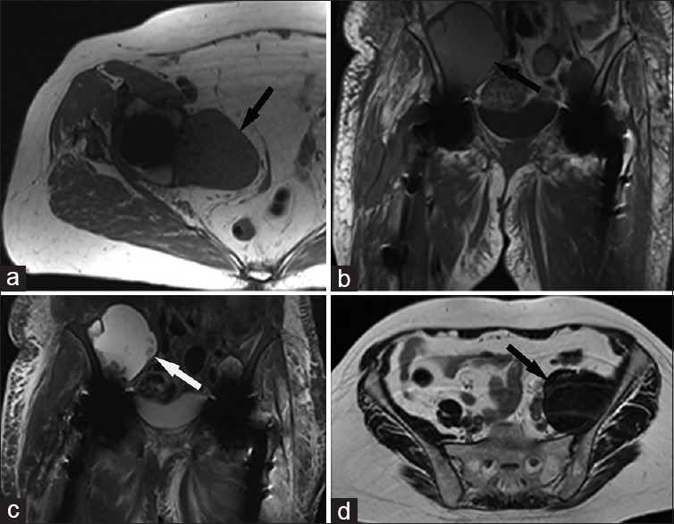
Figure 4.

75-year-old woman with a right total hip arthroplasty presents with a pelvic mass of unknown origin. (a) Axial T1-weighted magnetic resonance image of the right hip shows a T1 intermediate pelvic mass (arrow) which is isointense to skeletal muscle. 77-year-old female with a right iliopsoas mass and bilateral total hip arthroplasty. (b) Cor T1 and (c) short tau inversion recovery-weighted magnetic resonance images of the pelvis show a right-sided mass (arrows) which is hyperintense on T1 and T2. sixty-seven-year-old female with a left total hip arthroplasty and asymptomatic retroperitoneal mass. (d) Axial T2-weighted magnetic resonance image shows a large T2 dark mass (arrow) in the expected location of the left iliopsoas muscle.
