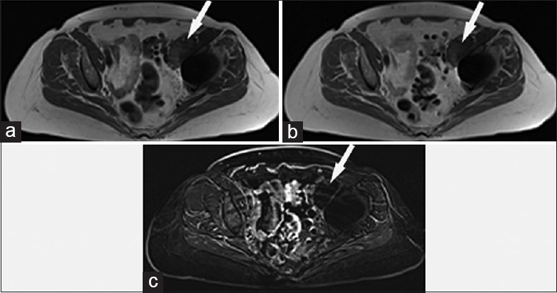
Figure 5.

68-year-old woman with a left total hip arthroplasty and concern for a malignant left-sided periarticular mass. (a) Axial T1-weighted magnetic resonance image shows a focal left-sided periarticular mass (arrow) which hyperintense to skeletal muscle. (b) Contrast-enhanced axial T1-weighted magnetic resonance image shows similar appearance to the noncontrast sequence, although the T1 hyperintense nature of the mass (arrow) limits diagnostic sensitivity for detection of enhancement. (c) Axial subtraction sequence magnetic resonance image (produced by subtraction of the pre- and post-T1-weighted sequences) shows no enhancement of the mass (arrow).
