
Figure 6.
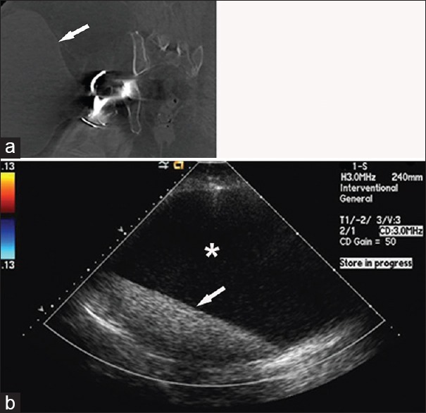
69-year-old woman with a right total hip arthroplasty presents with the right hip pain and swelling. (a) Contrast-enhanced coronal computed tomography image of the right hip shows a large cystic-appearing mass (arrow) associated with the right hip and extending into the subcutaneous soft tissues. (b) Duplex color Doppler ultrasound confirms the cystic nature of the mass. The mass is avascular, contains anechoic fluid (asterisk), and has layering internal echogenic debris (arrow).