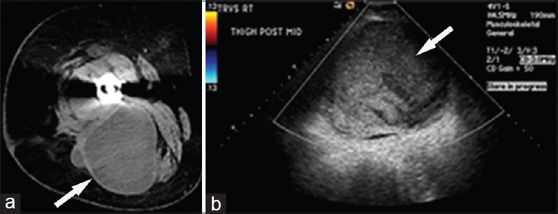
Figure 7.

69-year-old woman complains of on-going right hip pain following placement of a right total hip arthroplasty. (a) Contrast-enhanced axial computed tomography image of the right hip shows a large mixed density mass (arrow) in the posterior compartment of the right thigh. (b) Duplex color Doppler ultrasound shows an avascular mass (arrow) with mixed internal echogenicity and posterior acoustic enhancement.
