
FIGURE 4.
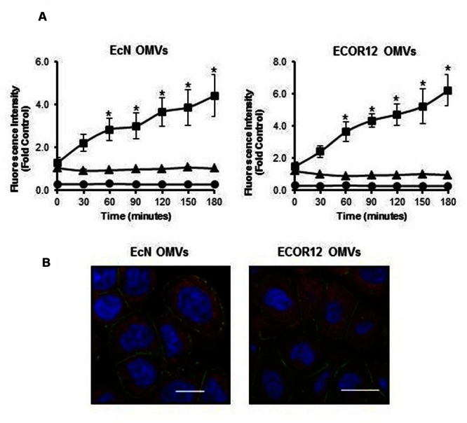
Outer membrane vesicles uptake by differentiated Caco-2 cells. (A) Rhodamine B-R18-labeled OMVs from EcN or ECOR12 were applied to the apical side of polarized Caco-2 cells and fluorescence was measured over time (squares). OMVs (triangles) and cells (circles) alone were used as negative controls for background fluorescence. Statistical differences versus the for background fluorescence emitted by OMVs alone were assessed by one-way ANOVA followed by Tukey’s test (∗p < 0.05). (B) Visualization of internalized OMVs by fluorescence microscopy. Caco-2 cells were incubated with rhodamine B-R18 labeled OMVs for 1 h at 37°C. The cell membrane was visualized by immunostaining with antibodies against the zonula occludens ZO-1 protein followed by Alexa Fluor 488-conjugated secondary antibody (green). Nuclei were stained with DAPI (blue). Internalized rhodamine-R18 labeled OMVs are visualized in red. Bar: 20 μm.