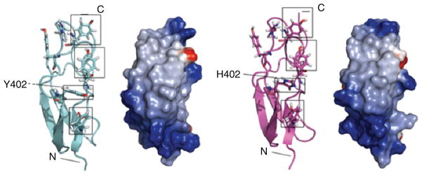
Fig. 3.
Comparison of structures of FH-7. Secondary structure and electrostatic surface representations (red = negative, blue = positive) are shown for the Y402 (left-hand panels) and H402 (right-hand panels) allotypic variants. Residue 402 is labeled and boxed and three tyrosine side-chains on the same face of the module are also boxed

