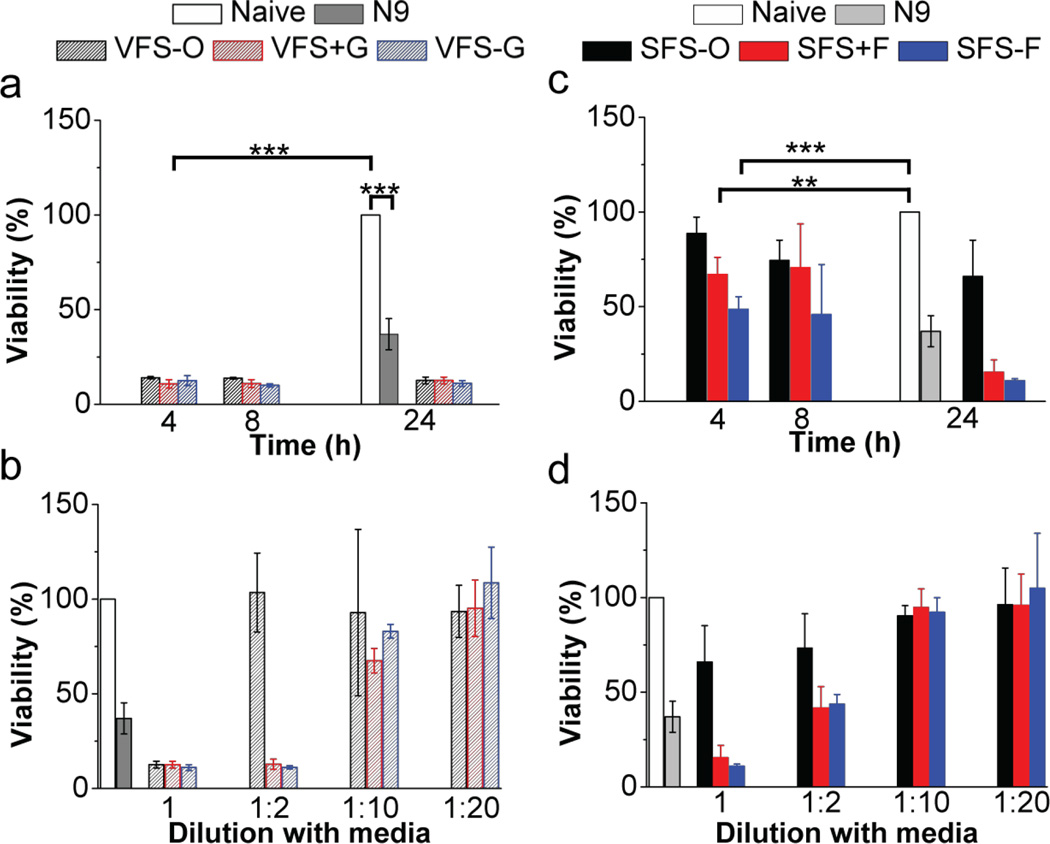
Figure 4.
Cytotoxicity of VFS and SFS buffers in HEC-1-A cells. Naïve cells and N9 (0.1 mg/mL; N = 3) were used as controls and effect of exposure time (4, 8 and 24 h) and dilution with cell culture media followed by 24 h dosing was studied (N = 5, Mean ±SD). N9 (p < 0.001; unpaired t-test with two-tailed distribution and unequal variance) and undiluted VF compositions (p < 0.001; single factor ANOVA) showed significant toxicity with respect to no treatment. An increase in viability with VFS compositions diluted with cell culture media in the ratio 1:2, 1:10 and 1:20 was noted. Whole SFS-O was not toxic and diluted SFS−/+F (1:10 and 1:20) showed enhanced cell viability with no significant difference from untreated cells (p > 0.05; single factor ANOVA).

