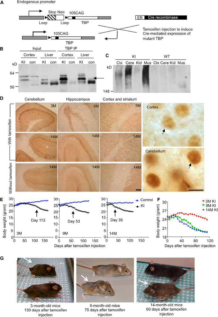
Figure 1. Characterization of TBP105Q Inducible KI Mouse Model.
(A) A schematic illustration of expression of mutant TBP in TBP105Q inducible KI mice.
(B) Western blotting analys is of immunoprecipitated TBP from the cortex and liver of WT and TBP105Q inducible KI mice after tamoxifen induction for two months. The rabbit polyclonal antibody EM192 was used to immunoprecipitate TBP, and the mouse monoclonal antibody 1C2 was used to visualize mutant TBP (arrow).
(C) Mutant TBP aggregates in the stacking gel were clearly detected in the cortex (Ctx), cerebellum (Cere), and muscle (Mus) of TBP105Q inducible KI mice by anti-TBP (1TBP18) but not in the kidney (Kid).
(D) 1C2 immunohistochemical study confirming mutant TBP expression in the cerebellum, hippocampus, cortex, and striatum of 3- and 14-month-old KI mice injected with tamoxifen. Small aggregates of mutant TBP were visualized in the nucleus under 1003 magnification (arrows). Scale bars represent 100 µm(left) and 5 µm (right).
(E) TBP105Q inducible KI and control mice at 3, 9, or 14 months of age were injected with tamoxifen in order to induce the expression of mutant TBP. The older KI mice showed earlier onset of body weight loss (day 35 for 14 months, p = 0.0374) than the younger KI mice (day 53 for 9 months, p = 0.0125, and day 113 for 3 months, p = 0.0438) after tamoxifen injection (n = 9–11 each group, two-way ANOVA, p < 0.0001).
(F) The body weight of differently aged KI mice was also compared.
(G) Representative images of differently aged (3, 9, and 14 months) KI mice (white arrow) were taken at the indicated days after tamoxifen injection.

