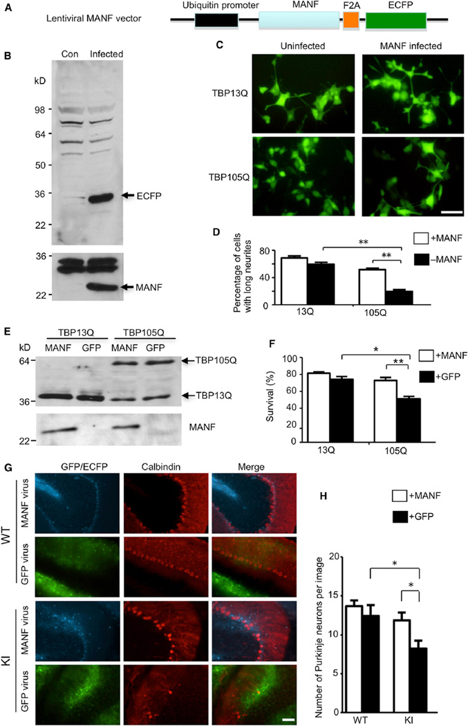
Figure 7. Overexpression of MANF Ameliorates Toxicity Caused by Mutant TBP In Vitro and In Vivo.
(A) Schematic map of lentiviral MANF vector.
(B) Western blotting analysis of HEK293 cells infected with lentiviral MANF for two days (Infected) confirming the expression of both MANF and ECFP. Uninfected HEK293 cells (Con) were used as a negative control.
(C and D) PC12 cells stably expressing either WT TBP (TBP13Q) or mutant TBP (TBP105Q) were infected with lentiviral MANF or uninfected. Fluorescent imaging (C) and quantitative analysis (D; n = 15, **p < 0.01) showed that MANF significantly increased the percentage of cells with long neurites. The scale bar represents 10 µm.
(E) Western blot analysis of PC12 cells stably expressing either WT TBP (TBP13Q) or mutant TBP (TBP105Q), which were infected with lentiviral MANF or GFP.
(F) MTS assay showed a significant increase in the survival of PC12 cells (versus non-staurosporine-treated cells) expressing mutant TBP when infected with lentiviral MANF in comparison to lentiviral GFP (*p < 0.05, **p < 0.01).
(G) One-year-old WT and TBP105Q inducible KI mice were injected with MANF lentivirus on the right side of the cerebellum and with lentiviral GFP on the left side. Fluorescence of GFP or ECFP was used to locate the region infected by lentivirus. Calbindin staining showed that MANF overexpression rescued the loss of Purkinje neurons caused by mutant TBP. The scale bar represents 50 µm.
(H) Quantitative assessment of the number of Purkinje cells labeled by anticalbindin per image field (20×, n = 9, *p < 0.05). Data are represented as mean ± SEM.

