Figure 4.

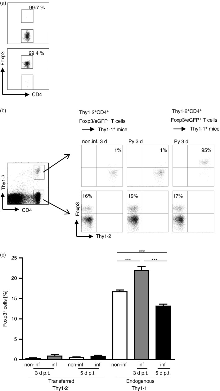
Adoptively transferred CD4+ Foxp3− T cells do not acquire Foxp3 expression in Plasmodium yoelii‐infected mice. From 3 × 10⁶ to 5 × 10⁶ sorted Thy1.2+ CD4+ Foxp3− T cells or Thy1.2+ CD4+ Foxp3+ T cells from naive Foxp3/eGFP mice were injected intravenously into Thy1.1+ BALB/c mice before infection with 1 × 10⁵ infected red blood cells (iRBC) or in naive, non‐infected mice. At 3 and 5 days post‐infection (d p.i.) mice were killed and Foxp3 expression was analysed in gated CD4+ Thy1.2+ and CD4+ Thy1.1+ T cells, respectively by flow cytometry. (a) Purity of sorted CD4+ Foxp3/eGFP− T cells and CD4+ Foxp3/eGFP+ T cells is shown in representative dot plots. (b) Representative dot plots for either non‐infected or P. yoelii‐infected Thy1.1 mice receiving either Thy1.2+ CD4+Foxp3− T cells or Thy1.2+ CD4+ Foxp3+ T cells 3 d p.i. / cell transfer. (c) Data from two or three independent experiments with n = 4 to n = 11 mice in total are summarized as mean ± SEM. Student's t‐test was used for statistical analysis. ***P < 0·001.
