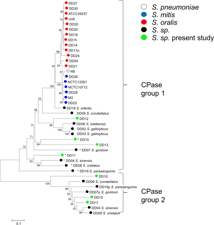
FIG 6 .
Distribution of PBP3 homologs in streptococcal genomes. A phylogenetic tree was constructed from deduced protein sequences from PBP3 d,d-carboxypeptidases using the MEGA6 software and muscle alignment. Proteins with at least one unusual active-site motif, indicating a nonfunctional PBP, are marked by asterisks, and partial sequences indicated by P bootstrap values (percentages) are based on 1,000 replications. The bar refers to genetic divergence as calculated by the MEGA software.

