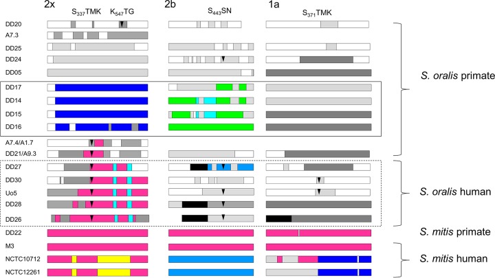
FIG 7 .
Mosaic PBPs in S. mitis and S. oralis. Mosaic gene structures were deduced by comparison to S. oralis ATCC 35037 PBP genes. Sequences that are highly similar to each other (<5% difference) are shown in the same color; sequences of different colors diverge from each other by at least 15%. Light gray, divergence from S. oralis ATCC 35037 of approximately 5%; dark gray, divergence by >15%; arrowheads, mutations within or close to active-site motifs which are shown on top; solid-line box, PBPs from free-living chimpanzees; dashed box, PBPs from human isolates with high-level penicillin resistance. The amino acid numbers are those of PBPs from sensitive S. pneumoniae strains.

