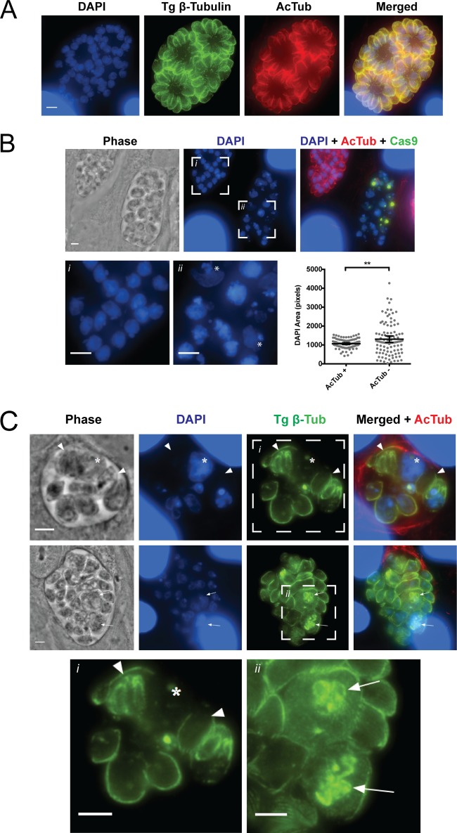
FIG 6 .
Defects in nuclear division and segregation in parasites lacking α-tubulin K40 acetylation. (A) TgATATHA parasites were transfected with GFP-Cas9-sgTgATAT and imaged at 40 h posttransfection. Shown are transfectants lacking GFP-Cas9 expression, which display normal replication and have microtubules containing K40 acetylation, as visualized with anti-β-tubulin (green) and acetyl-K40–α-tubulin (red) antibodies, with DAPI costain in blue. (B) Parasites expressing GFP-Cas9 lose K40 acetylation and contain abnormal nuclear morphology compared to that of parasites possessing K40 acetylation. Nuclei were visualized by DAPI staining (blue). Insets of acetyl-K40-positive (i) and -negative (ii) parasites are shown and expanded in the lower panels. The DAPI-stained structures was measured with ImageJ (n = 100 nuclei). Double asterisks indicate a significant difference in mean area, as determined by unpaired t test with Welch’s correction for unequal variance (P = 0.0085). (C) Vacuoles containing parasites lacking acetylated microtubules (red) and showing aberrant phenotypes detected by staining all of the microtubules (green) and DNA (blue) are shown. Inset i shows that parasites lacking K40 acetylation have defects in microtubule structures and fail to partition nuclear material into daughter parasites. Anucleate parasites are marked by arrowheads, while the improperly segregated nuclear mass is indicated by the asterisk. Inset ii shows parasites containing multiple β-tubulin structures resembling daughter cell conoids (arrows). Scale bar, 3 μm.

