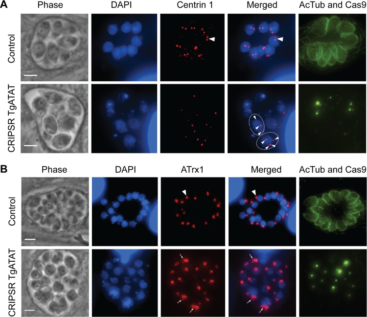
FIG 7 .
Centrosome duplication and apicoplast division in parasites lacking K40 acetylation. (A) Duplication of the centrosomes occurs in the presence (top row, arrowhead) or absence of K40 α-tubulin acetylation (bottom row, arrowheads), as visualized by IFA staining for centrin 1 (red), acetyl-K40–α-tubulin (green), and DNA (DAPI, blue). K40-acetylated α-tubulin and GFP-Cas9 (localized to the nucleus) were detected in the same channel (green). Note the loss of acetylated α-tubulin and GFP-Cas9-expressing parasites. Dotted lines encircle individual parasites with arrowheads indicating multiple centrosomes. (B) IFAs of parasites stained for apicoplast membrane protein Atrx1 (red), acetyl-K40–α-tubulin (green), DNA (DAPI, blue), and GFP-Cas9 (green, nuclear). Apicoplasts that underwent normal division are visible in acetyl-K40-positive parasites (top row, arrowhead). Apicoplasts that failed to divide in parasites lacking K40 acetylation (bottom row) are indicated by arrows. Scale bars, 3 μm.

