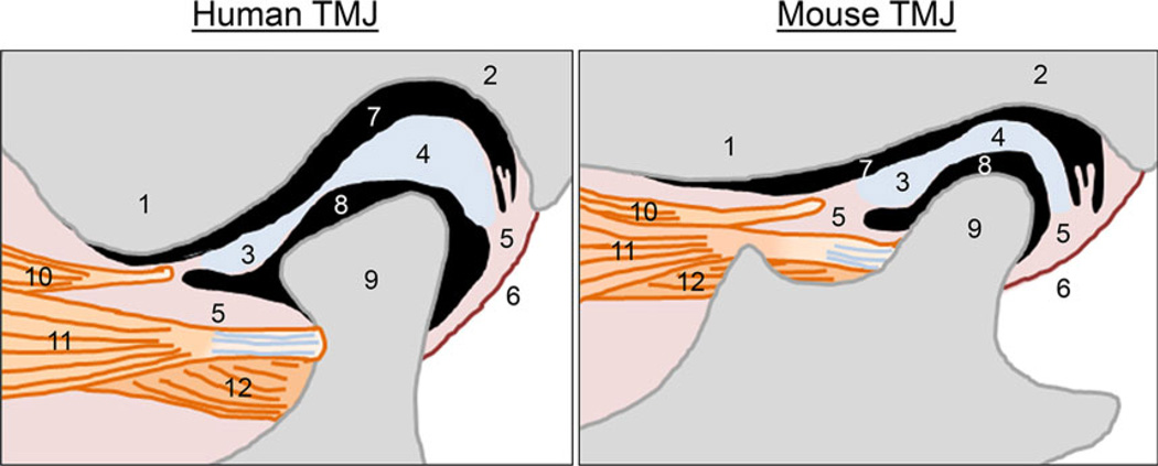
Figure 2.
Comparison of the structure of the temporomandibular joint between humans and mice. TMJ, temporomandibular joint. 1: the articular eminence of the temporal bone, 2: the glenoid fossa of the temporal bone, 3: anterior band of the articular disk, 4: posterior band of the articular disk, 5: connective tissue, 6: the posterior joint capsule, 7: the upper articular cavity, 8: the lower articular cavity, 9: mandibular condyle, 10: a part of upper head of the lateral pterygoid muscle, associated with the articular disk, 11: upper head of the lateral pterygoid muscle, connected with the mandibular condyle, 12: lower head of the lateral pterygoid muscle, connected with the mandibular condyle

