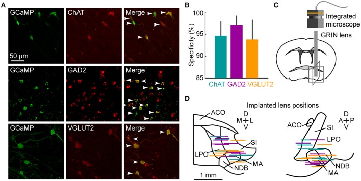
Figure 1.
Microendoscopic calcium imaging in the BF. (A) Examples of GCaMP6f (green) and cell-type-specific markers (red: ChAT, immunohistochemical staining; GAD2 and VGLUT2, fluorescence in situ hybridization) expressed in the respective Cre mice. Arrowheads mark double-labeled neurons. (B) Specificity of GCaMP6f expression. 94.4 ± 1.7% of 399 GCaMP6f+ neurons in 5 ChAT-Cre mice; 96.2 ± 1.3% of 879 neurons in 7 GAD2-Cre mice and 93.6 ± 2.4% of 770 neurons in 5 VGLUT2-Cre mice were also labeled by the cell-type-specific marker. Data are presented as mean ± SEM. (C) Schematic of microendoscopic imaging in BF. Gray boxes correspond to fields of view in (D). (D) Placement of GRIN lenses in the BF, viewed in the coronal (left) and sagittal (right) planes. Each colored line indicates the position of the bottom surface of the lens in one animal. There were no significant differences in implanted lens position between ChAT-, GAD2- and VGLUT2-Cre mice (Fgenotype = 0.004, p = 0.99, 2-way repeated measures (RM) ANOVA, n = 9 ChAT, 7 GAD2, 7 VGLUT2 mice). ACO, anterior commissure; LPO, lateral preoptic area; NDB, diagonal band nucleus; MA, magnocellular area; SI, substantia innominata.

