Figure 2.
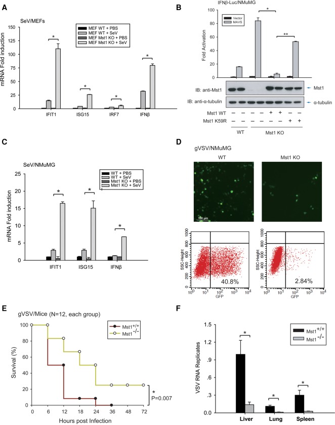
Mst1 negatively regulates host antiviral defense in primary/cultured cells and mice. (A) Antiviral response of control or Mst1−/− MEFs against SeV infection was measured by mRNA induction at 12 hpi of various ISGs, including IFIT1, ISG15, IRF7, and IFNβ. Mst1−/− MEFs exhibited stronger antiviral responses than control MEFs. (*) P < 0.05, compared with control MEFs, by Student's t-test. (B, bottom panel) Mst1−/− NMuMG epithelial cells were generated by the CRISPR/Cas9 method, and the Mst1 knockout (KO) and rescue were verified by immunoblotting. Cytosolic RNA-sensing signaling in Mst1−/− and control NMuMG was evaluated by IFNβ reporter assay. (Top panel) Stronger IRF3 transactivation was observed by Mst1 ablation but was reversed by the reintroduction of wild-type (WT) Mst1. (C) Antiviral response in Mst1−/− and control NMuMG cells to SeV infection at 6 hpi was measured by quantification of ISGs and IFNβ, and stronger antiviral responses were observed by Mst1 deletion in NMuMG cells. n = 4 Mst1−/− clones. (*) P < 0.01, compared with control NMuMG cells, by Student's t-test. (D) Mst1−/− and control NMuMG cells were infected with GFP-tagged vesicular stomatitis virus (gVSV). Reduced virus replication, as evidenced by a reduced level of GFP+ cells, was observed in Mst1−/− NMuMG cells by microscopy (top panels) or FACS assay (bottom panels). (E) Survival of ∼8-wk-old Mst1+/+ and Mst1−/− mice given intravenous tail injection of gVSV (2 × 10⁷ plaque-forming units [pfu] per gram). n = 12 mice for each group. P < 0.01, by paired Student t-test. (F) Determination of gVSV loads in mouse organs at 12 hpi in Mst1+/+ and Mst1−/− mice, which were intravenously tail-injected, with gVSV. n = 3 mice for each group. (*) P < 0.01, compared with control Mst1+/+ group, by Student t-test.

