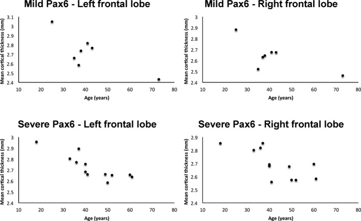
Figure 6.

Results of region‐of‐interest analysis showing that cortical thickness declines significantly with age in the frontal lobes in severely affected PAX6 subjects (bottom row), but not in the mildly affected PAX6 subjects (top row). Parameter assessments and partial correlations, with a Bonferroni‐corrected alpha of 0.004, show that while controls and mildly affected PAX6 subjects had no correlation between left or right frontal lobe thickness and age, respectively (top row), a borderline significant negative correlation was present for severely affected PAX6 subjects (bottom row) (r = −0.857, P < 0.001 and r = −0.755, P = 0.004).
