
Figure 4.
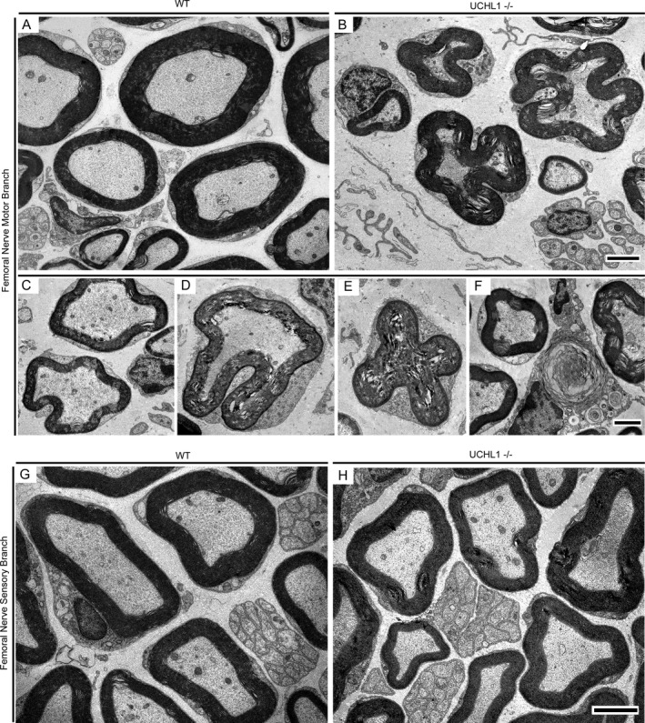
Axon fibers in the motor branch of femoral nerve display selective axonal degeneration. (A) A representative cross section image of axons within the motor branch of the femoral nerve in WT mice. Axons with different sizes, and the intact myelin sheath around them is observed with electron microscope. (B) A representative cross‐sectional image of axons within the motor branch of the femoral nerve in UCHL1−/− mice. Axons fibers with various degrees of degeneration are present. (C‐E) Representative images displaying stages of axonal defects. (C) Initial stage of axon fiber collapse, (D) intermediary stage showing invagination of myelin sheath, and (E) a completely collapsed axon fiber. (F) An example of completely degenerated axon fiber being scavenged by macrophage. (G‐H) Representative cross‐sectional electron micrographs of sensory branch of the femoral nerve in WT (G) and UCHL1−/− (H) mice appear comparable, and both are devoid of major axonal defects. Scale bar: 2 μm.