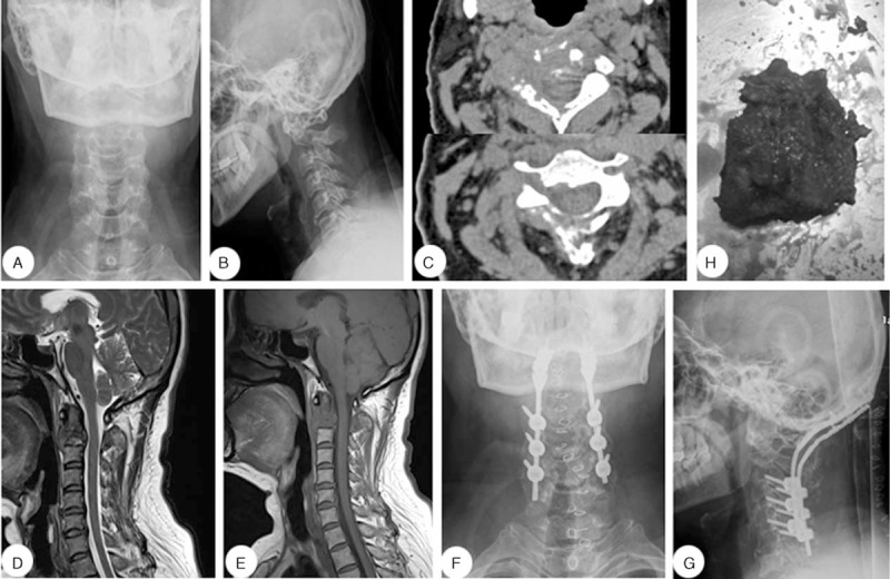
FIGURE 1.

A 51-y-old woman diagnosed with a C2 vertebral metastasis. (A, B) Preoperative lateral and anteroposterior cervical X-rays. (C) Preoperative computed tomography scan. (D, E) Preoperative sagittal T1- and T2-weighted magnetic resonance imaging scans. (F, G) Postoperative lateral and anteroposterior cervical X-ray images.
