
Figure 2.
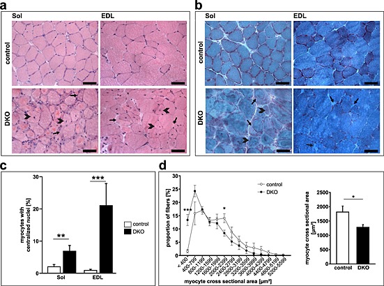
Absence of muscle‐specific RING‐finger (MuRF)2 and MuRF3 resulted in protein surplus myopathy. (A) Haematoxyline and eosin stain of cross‐sections from soleus (Sol) and extensor digitorum longus (EDL) from 21‐week‐old control and DKO mice. Subsarcolemmal accumulations of eosinophilic material occurred around a central core of myofibres in DKO muscles (arrow). Heterogeneity of fibre size and centrally localized nuclei (arrowhead) were found in DKO muscles. Scale bar, 50 µm. (B) Gomori's trichrome stain of cross‐sections from Sol and EDL from 18‐ to 21‐week‐old control and DKO mice. Protein aggregates (arrow) and myofibres with centralized nuclei are depicted (arrowhead). (C) Quantification of myofibres containing centralized nuclei in Sol and EDL of 20‐ to 22‐week‐old control (n = 13) and DKO (n = 8) mice. **P < 0.01, ***P < 0.001. (D) Quantification of myocyte cross‐sectional area (MCSA) from control (n = 7) and DKO (n = 7) mice on cross‐sections from EDL. An increased number of smaller fibres were found in DKO. *P < 0.05, ***P < 0.001.