
Figure 3.
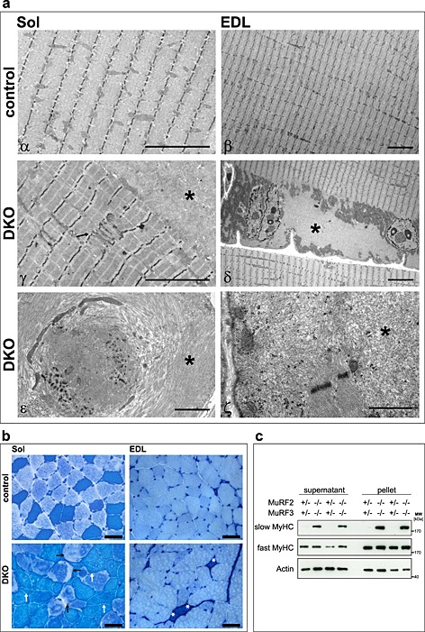
Double knockout (DKO) mice displayed a protein aggregate myopathy and a shift towards slow myofibres in extensor digitorum longus (EDL) muscle. (A) Electron microscopy of soleus (Sol) and EDL sections displayed accumulation of amorphous material in DKO myofibres. Asterisks indicate accumulating aggregates, arrow points to abnormal Z‐line. Scale bar, 5 µm (α–δ), 2 µm (ε, ζ). (B) ATPase stain of Sol and EDL showed accumulations inside of slow/type I fibres (dark blue, white arrows) and fast/type II fibres (bright blue, black arrows) of DKO mice. Slow/type I fibres occurred in EDL of DKO mice (asterisk). Scale bar, 50 µm. (C) Immunoblotting of proteins from the soluble (supernatant) and particulate (pellet) fractions of EDL revealed accumulation of slow/type I myosin heavy chain protein in DKO muscle. Actin was used as a control.