
Figure 4.
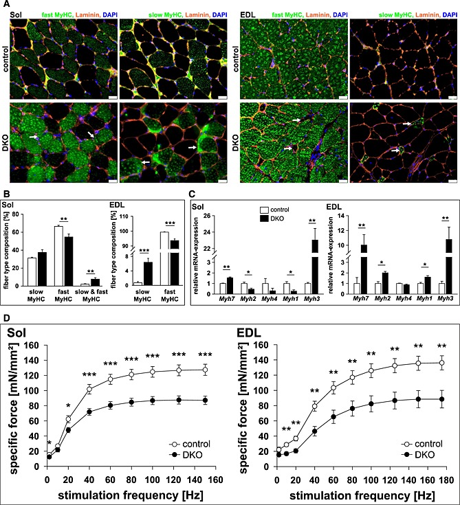
Loss of muscle‐specific RING‐finger (MuRF)2 and MuRF3 leads to an increase in slow/type I fibres in skeletal muscle. (A) Immunohistochemistry of cross‐sections from soleus (Sol, left panel) and extensor digitorum longus (EDL, right panel) using anti‐laminin, anti‐fast/type II myosin heavy chain, or anti‐slow/type I myosin heavy chain antibody. Nuclei were stained with 4',6‐Diamidino‐2‐phenylindol (DAPI). Protein aggregations are indicated (arrows). Slow/type I fibres occurred in EDL of DKO mice (arrow). Scale bar, 25 µm. (B) Slow/type I and fast/type II MyHC containing fibres were quantified in Sol and EDL of control (n = 8–10) and DKO (n = 7–8) mice. Data are shown as mean ± SEM. **P < 0.01, ***P < 0.001. (C) Real‐time RT–PCR analysis of myosin heavy chain (Myh) 1, 2, 3, 4 and 7 gene expression in Sol and EDL from control (n = 8–9) and DKO (n = 4) mice. Hypoxanthine guanine phosphoribosyl transferase (Hprt) expression was used as reference. Data are presented as mean ± SEM. *P < 0.05, **P < 0.01. (D) Maximal force development of Sol and EDL from male 14‐ to 23‐week‐old control (n = 9 each) and DKO (n = 11–12) mice is shown. Specific force [mN/mm2] per stimulation frequency [Hz] is depicted. Data are presented as mean ± SEM. *P < 0.05, **P < 0.01, ***P < 0.001.