
Figure 9.
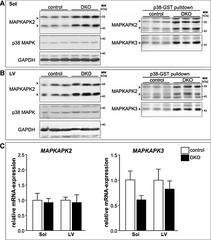
Mitogen‐activated protein kinase‐activated protein kinase (MAPKAPK) 2 and MAPKAPK3 are increased in double knockout (DKO) soleus (Sol) and left ventricle. (A) Immunoblotting of proteins from total tissue lysates of Sol from control and DKO mice using anti‐MAPKAPK2 and anti‐p38 MAPK antibody. GAPDH served as loading control (left panel). GST‐p38 MAPK pulldown assays were performed with tissue lysates from soleus of control and DKO mice and analysed using anti‐MAPKAPK2 antibody and anti‐MAPKAPK3 antiserum (right panel). (B) Immunoblotting of proteins from total tissue lysates of left ventricles (LV) from control and DKO mice using anti‐MAPKAPK2 and anti‐p38 MAPK antibody. GAPDH served as loading control (left panel). GST‐p38 pulldown assays were performed with tissue lysates from hearts of control and DKO mice and analysed using anti‐MAPKAPK2 antibody and anti‐MAPKAPK3 antiserum (right panel). (C) Real‐time RT–PCR analysis of MAPKAPK2 and MAPKAPK3 gene expression in Sol and LV from control (n = 8–9) and DKO (n = 4) mice. Hypoxanthine guanine phosphoribosyl transferase (Hprt) expression was used as reference. Data are presented as mean ± SEM.