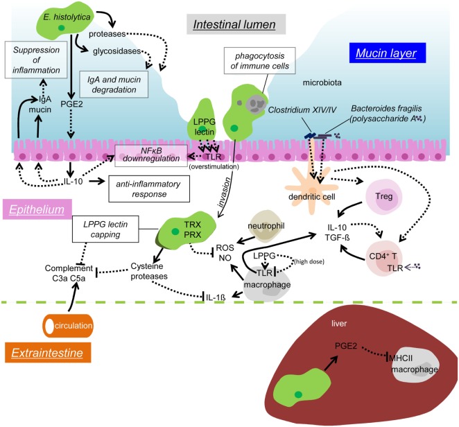
Figure 2.
Possible mechanisms of immune evasion during amebiasis. Secreted or surface proteases of the amebae degrade IgA in the mucosal layer. PGE2 from the amebae induces IL-10 secretion from the IECs, and in turn stimulates mucin and IgA secretion, which likely prevents unnecessary inflammation. Overstimulation of TLR causes downregulation of NFκB activation. Removal of infiltrating immune cells by phagocytosis/trogocytosis helps to reduce immune responses. Some commensal microbiota, namely Clostridium XIV and IV groups and Bacteroides fragilis, induce Treg cells to downregulate immune responses. Polysaccharide A from B. fragilis binds to TLR2 on CD4 T cells and induces IL-10 production. The amebae in the tissues and the blood stream evade from complement by surface receptor capping (LPPG, lectin) and degradation of C3a and C5a by cysteine proteases. Cysteine proteases also degrade IL-1β, antioxidative stress defense by the TRX and PRX systems fends off the attack from ROS and NO from activated neutrophils and macrophages. LPPG binds to TLR2 on monocytes and macrophages, which leads to secretion of cytokines, including IL-10 and TGF-β. High doses of LPPG downregulate TLR2 gene expression in monocyte and cause negative feedback of protective immune responses. PGE2 from the amebae and the host causes downregulation of MHC class II expression on macrophages in the liver, which results in anti-inflammation.

