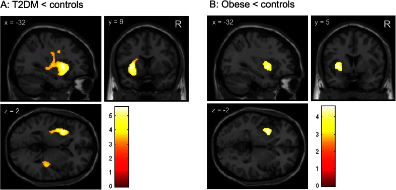
Fig. 2.
Reduced white matter volume in T2DM patients compared with lean subjects. a Brain slices showing clusters of reduced white matter volume in the external capsule region in obese T2DM patients compared with lean subjects, as determined with VBM; b Brain slices showing a cluster of reduced white matter volume in the external capsule region in obese compared with lean subjects. This cluster, however, was not statistically significant after FWE-correction for multiple comparisons (PFWE = 0.380). The color scale reflects the T-value. Right side of the axial slices is the right side of the brain. X, y, z are the Montreal Neurological Institute (MNI) coordinates of the brain in standard space

