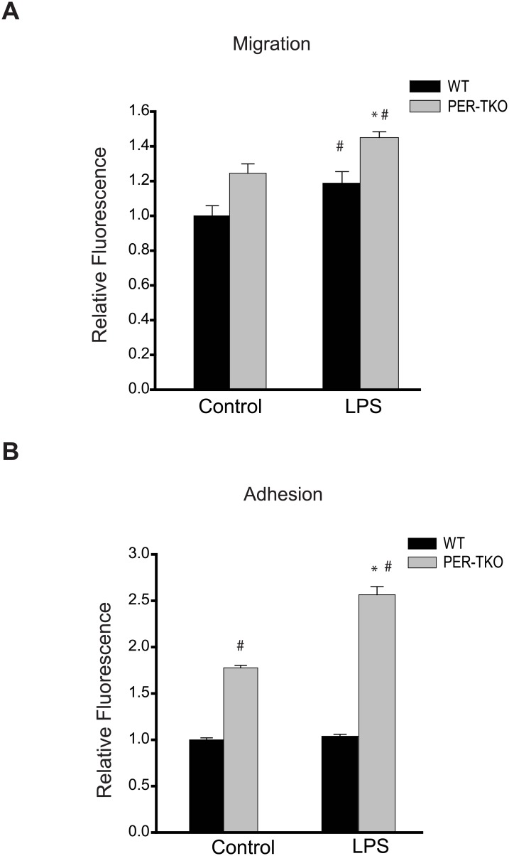
Fig 8. Circadian clock disruption alters the migration and adhesion of macrophages.
(A) The effect of circadian disruption on macrophage migration was evaluated using the Oris Cell Migration Assay. Peritoneal macrophages were isolated from WT and Per-TKO mice and subjected to the indicated treatment (LPS 100ng/ml) for 24h, cells stained with calcein AM for 30 min and the fluorescence signal in the migration zone quantified. (mean ± SEM n = 4–5, two-way ANOVA with Bonferroni post hoc correction, *p<0.05 versus control, # p<0.05 versus WT). (B) Fluorescently labeled peritoneal macrophages were incubated with activated adherent human aortic endothelial cells for 15 minutes at 37°C and the degree of cell adhesion assay was quantified (mean ± SEM n = 6, two-way ANOVA with Bonferroni post hoc correction, *p<0.05 versus control, # p<0.05 versus WT).

