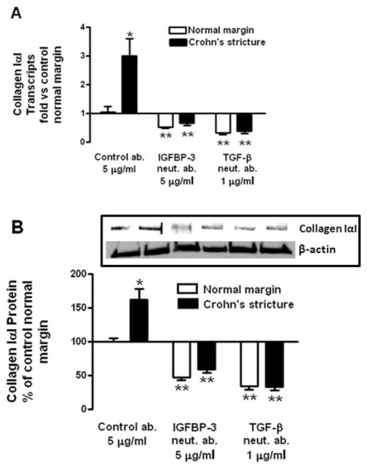
FIGURE 6.
Endogenous IGFBP-3 and TGF-β1 regulate excess collagen IαI expression and production in normal and stricturing muscle of CD. (A) Immunoneutralization of endogenous IGFBP-3 and TGF-β1 decreases collagen IαI expression in both normal margin muscle cells and in muscle cells isolated from strictures in CD. Collagen IαI RNA transcript levels were measured by qRT-PCR. Results are expressed as fold versus basal normal margin muscle cells using the 2−ΔΔCt method with β-actin as control. (B) Immunoneutralization of endogenous IGFBP-3 and TGF-β1 decreases collagen IαI protein production in both normal margin muscle cells and in muscle cells isolated from strictures of CD. Inset: representative immunoblots of collagen IαI and β-actin. Quiescent muscle cells were incubated for 24 hours with preimmune serum (control ab.), or neutralizing antibodies to IGFBP-3 or TGF-β1. Collagen IαI protein levels were measured in conditioned media by immunoblot analysis. Results are expressed as percent of control normal margin muscle cells. Values represent mean ± SEM of 4–5 separate experiments. *P < 0.05 versus normal margin; **P < 0.05 versus control antibody-treated cells.

