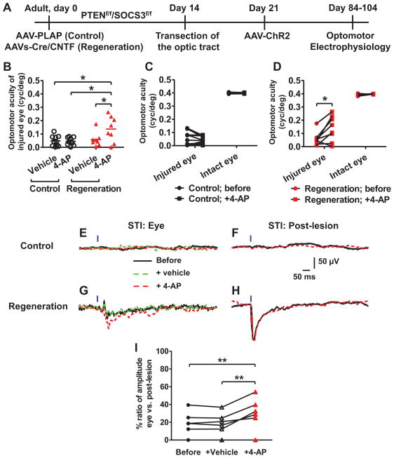
Figure 5. Improved optomotor acuities achieved by combined treatment of 4-AP and axon regeneration in adult-injured mice.
(A) Schematic diagram showing the experimental timeline. Control group: PTENf/f/SOCS3f/f with AAV-PLAP and regeneration group: PTENf/f/SOCS3f/f with AAVs-Cre/CNTF. Mice of 8 weeks or older are considered as adults. (B–D) Optomotor acuities in injured and intact eyes of both groups. 4-AP treatment increased the optomotor acuities of injured eyes only in the regeneration group but not in control group (B). In the control group, 4-AP did not change the acuities of either injured or intact eyes (C). However, in the regeneration group, 4-AP increased the optomotor acuities of only injured eyes, but not intact ones (D). N = 10 for control and 8 for regeneration. (E–H) Representative eye-evoked (E, G) or post-lesion terminal-evoked (F, H) LFPs in control (E, F) or regeneration (G, H) group. Post-behavioral in vivo recordings showed 4-AP, but not vehicle, significantly increased eye-evoked LFPs in regeneration-group mice. No LFPs were recorded in all control-injured mice with or without 4-AP. (I) Ratios of eye-evoked vs paired terminal-evoked LFPs showing the effect of 4-AP or vehicle in regeneration-group mice. N = 7 (out of 8; one failed recording due to technical reasons). No eye-evoked LFPs were recorded in two of them. * P < 0.05, ** P < 0.01 ANOVA with Bonferroni post-test. See also Figure S5.

