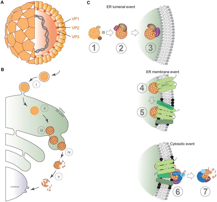
Figure 3. The non-enveloped virus SV40 co-opts elements of the ERAD machinery to translocate into the cytosol.
A. Structure of SV40. SV40 consists of 72 homo-pentamers formed by the coat proteins VP1, with each pentamer encasing either a VP2 or VP3 internal protein (see cross-section). Within this proteinaceous shell, the viral circular double-stranded DNA genome is enclosed. B. SV40 infection pathway. To infect cells, the virus recognizes the host surface glycolipid (GM1) receptor. The virus-receptor complex undergoes endocytosis (step i) to reach the ER (step ii). In the ER lumen, several ER-resident factors isomerizes/reduces the virus disulfide bonds to generate conformationally-altered hydrophobic particles (step iii). These viral particles penetrate the ER membrane to reach cytosol with the aid of several ER-membrane and cytosolic factors (step iv). During cytosolic extraction, the virus is simultaneously disassembled (step v) to prepare a subviral intermediate (containing the genome) for transport into the nucleus for infection. C. Detailed overview of SV40 ER-to-cytosol transport. Step 1: Once SV40 reaches the ER lumen, ER-resident PDI family members including ERp57, ERdj5, and PDI act on the viral particle to trigger critical conformation changes. These structural rearrangements expose the internal proteins VP2 and VP3, generating a hydrophobic viral particle. Step 2: The hydrophobic viral particle recruits BiP via the action of the J-protein ERdj3. Step 3: The NEF Grp170 induces BiP release from SV40 proximal to the ER membrane. Step 4: The hydrophobic viral particle initiates membrane penetration by interacting with and integrating into the ER membrane via engaging the membrane protein BAP31. Step 5: BAP31 in turn recruits other membrane proteins (including the ER membrane J-proteins) to the vicinity to assemble a dense platform within the ER membrane (called foci, black dots) essential for membrane penetration. Step 6: At the cytosolic interface, several cytosolic factors (including Hsc70, SGTA, and Hsp105) anchored to the ER membrane engage the viral particle, extracting it into the cytosol in an energy dependent manner. Step 7: During cytosolic extraction, the virus is simultaneously disassembled to prepare for its journey into the nucleus.

