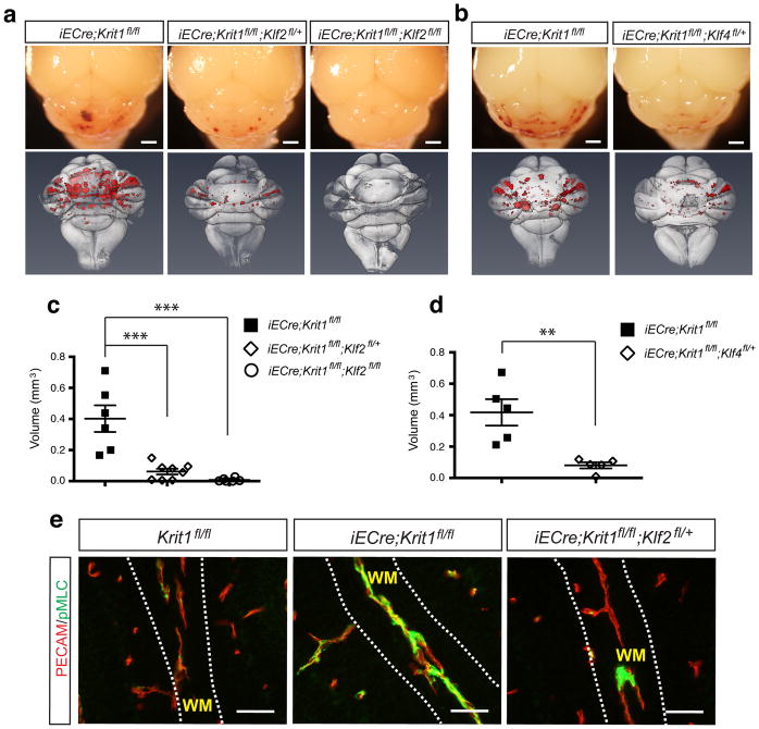
Figure 3. Genetic rescue of CCM formation and increased Rho activity with loss of KLF2 and KLF4.
a–b, Visual appearance of CCM lesions in Krit1ECKO, Klf2HetRSQ, Klf2HomoRSQ and Klf4HetRSQ animals is shown above and composite microCT images of the same hindbrains shown below. Scale bars, 1mm. c–d, Volumetric quantitation of CCM lesions in Krit1ECKO littermates with and without endothelial loss of one or two Klf2 alleles (c) or one Klf4 allele (d). *** indicates P<0.001; ** indicates P<0.01. N=5–7. e, Rescue of increased Rho activity by loss of KLF2. PECAM and pMLC staining of white matter (“WM”) vessels in the indicated P6 littermate brains is shown. Images are representative of 4 independent studies for each genotype. Scale bars, 50 μm.

