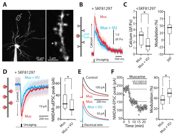
Figure 4. M4R activation on dSPNs inhibits postsynaptic NMDAR-mediated Ca2+ transients and currents.
(A) Low (left) and high (right) magnification maximum-intensity projections of a dSPN filled with Alexa Fluor 568. Spine was stimulated with 1 ms uncaging pulse of 720–725 nm light. (B) In the presence of the D1R agonist SKF81297 (3 μM), average NMDAR Ca2+ transients in a distal dSPN spine induced by a single glutamate uncaging pulse were reduced by the M4R PAM VU10010 (5 μM). Solid lines are averages across multiple spines and the shaded areas represent the mean ± SEM. (C) Box plot summary of modulation of NMDAR-mediated Ca2+ transients. (D) Addition of VU 10010 (5 μM) suppressed NMDAR-mediated uEPSC currents triggered by uncaging pulses of 500 Hz to five distal spines. Solid lines are averages across multiple spines and the shaded areas represent the mean ± SEM. (E) NMDAR-mediated EPSCs recorded from dSPNs in the presence of muscarine (3 μM; upper traces) and muscarine + VU10010 (5 μM; lower traces). EPSCs are averages of 10 consecutive trials. (F) Left: time course of EPSC amplitude from the experiment shown in (E). Muscarine and VU10010 were applied during the time indicated by the horizontal bars. Right: box plot sum of M4R mediated modulation of NMDAR currents. All box plot boxes indicate upper and lower quartiles; whiskers specify upper and lower 90%. *p < 0.05 (see also Figure S3).

