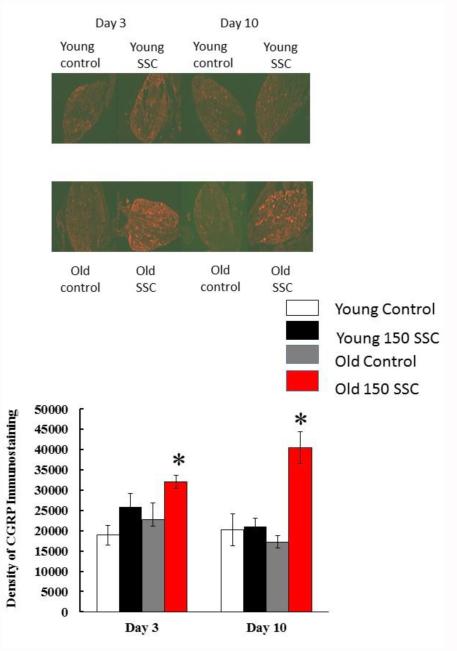
Figure 5.
The photomicrographs in (A) display the average density of CGRP staining in the ipsilateral DRG of young and old rats. Three days after iSSC exposure, staining density appeared to be higher in the DRG of injured as compared to young rats. However, this difference was only significant in old rats. Ten days after iSSC exposure,CGRP staining was still increased in the LTA of old rats exposed to SSCs. (*p < 0.05, different that time matched controls).

