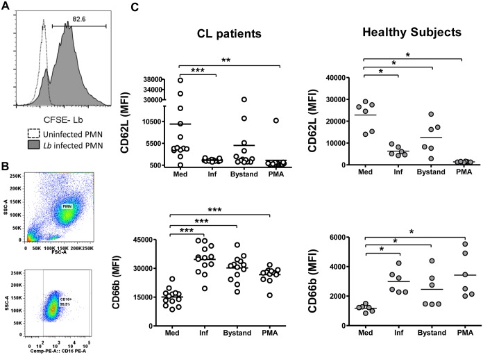
Fig 2. Effect of L. braziliensis infection on expression of neutrophil activation markers CD62L and CD66b.
Panel (A) shows a histogram demonstrating staining in neutrophils infected with CFSE-stained L. braziliensis versus uninfected neutrophils from a CL patient. Panel (B) shows a representative scatter plot indicating the purity of PMN population based CD16+ expression. Panel (C) shows collated results of surface staining for activation markers CD62L and CD66b by flow cytometry. Each value represents the MFI of one subjects’ neutrophils. “Inf” and “Bystand” represent the staining on neutrophils that were either infected (CFSE+) with CFSE- labeled L. braziliensis, or uninfected bystander neutrophils (CFSE-). The two left graphs represent data from subjects with CL; the two graphs on the right show data from healthy control subjects. Statistical analysis was performed using the Wilcoxon test, comparing stimulated to unstimulated cells (*p<0.05, **p<0.01, ***p<0.001).

