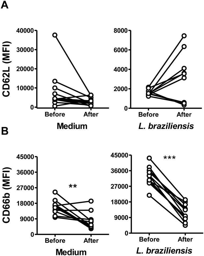
Fig 6. Expression of neutrophil surface markers CD62L and CD66b from CL patients before or after therapeutic cure.
Neutrophils from CL patients (n = 11) were isolated before or after CL treatment, and incubated with CFSE stained L. braziliensis at a 5:1 ratio. After 90 minutes cells were stained for flow cytometry and the expression of CD62L (A) and CD66b (B) was assessed by flow cytometry. Points on graphs represent the median MFI for neutrophils from each subject, incubated in medium alone or with CFSE labeled L. braziliensis. Statistical analyses utilized Wilcoxon test (**p<0.01, ***p<0.001).

