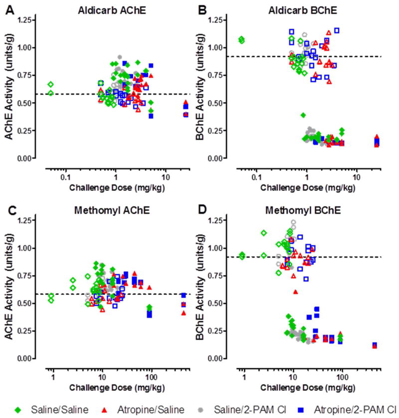
Figure 4. Terminal AChE and BChE activity in the cerebral cortex. AChE is unaffected by either aldicarb (A) or methomyl (C), however BChE activity is significantly reduced by both aldicarb (B) and methomyl (D).
Upon death due to challenge or at the completion of the 24hr observation period, the cortex was removed and ChE analysis was performed using an Ellman’s based assay. Closed circles represent animals that succumbed to challenge prior to 24 hours post-challenge. Open circles represent those animals that were euthanized following the 24h observation period. The dashed line is the average of all untreated control animals, n = 36. AChE/BChE activity is recorded in enzyme units per gram of tissue.

