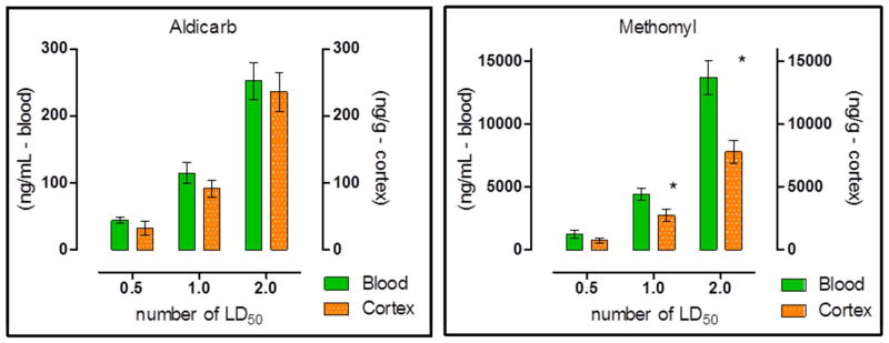
Figure 5. Blood and cerebral cortex concentration of Aldicarb (A) or Methomyl (B) 30 min post exposure.
Cerebral cortex was perfused prior to removal to prevent potential cross contamination between the blood and cortex. (Control non-perfused cortexes were collected to determine the impact of this potential cross contamination by the blood in the tissue found that no significate impact was present.) Animals were challenged with multiples of the previously determined MLD: 0.5, 1 or 2. Data shown are mean ± SEM, representing n = 5, (*) p<0.05, significance reflects difference between levels found in the blood and brain at a particular challenge level. Blood concentration is recorded in nanograms of carbamate per milliliter of blood, and cerebral cortex concentration is recorded in nanograms of carbamate per gram of tissue.

