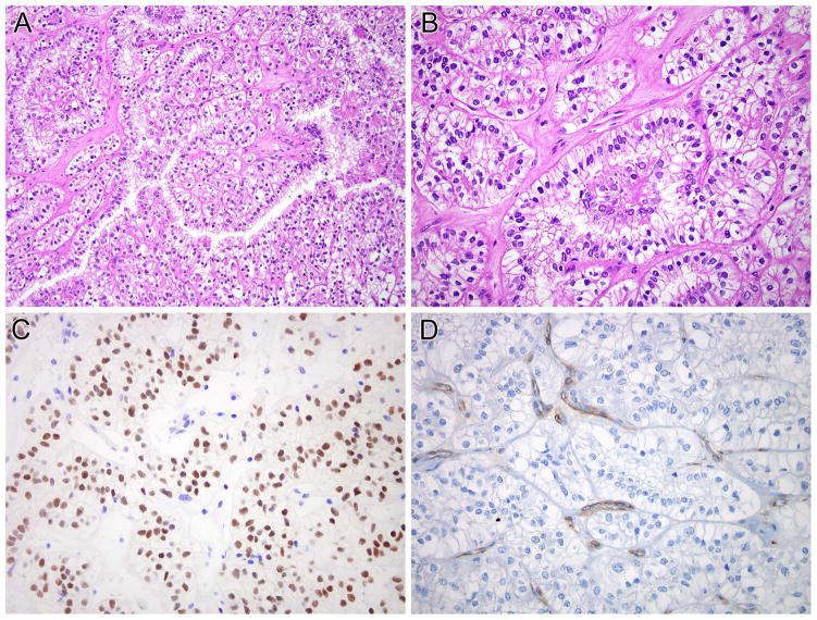
Figure 3.
NONO-TFE3 RCC (Table 1, case 4). A and B, this neoplasm demonstrated nested to papillary architecture. The neoplastic cells have predominantly clear cytoplasm and demonstrate sub-nuclear vacuolization, leading to apical palisading of nuclei. The neoplastic cells demonstrate nuclear labeling for PAX8 (C) but not for cathepsin K (D). Note the intact labeling of endothelial cells as an internal control for cathepsin K labeling.

