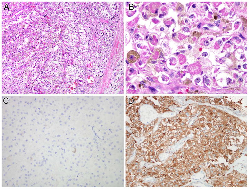
Figure 4.
NONO-TFE3 melanotic PEComa (Table 1, case 6). A and B, this is a neoplasm with nested to alveolar architecture, which features epithelioid cells with clear to finely granular eosinophilic cytoplasm. Fine pigment which proves to be melanin is present in the cytoplasm. The neoplasm was immunoreactive for melan A (not shown). The neoplasm does not label for PAX8 (C) but shows diffuse immunoreactivity for cathepsin K (D).

