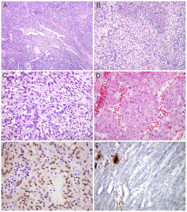
Figure 5.
DVL2-TFE3 RCC. This neoplasm demonstrates a variety of morphologic patterns. Much of the neoplasm has basophilic to pale cytoplasm, and demonstrates tubular and papillary architecture (A) each merges with sarcomatoid areas (B, C). Other areas on the same slides demonstrated more oncocytic cytoplasm (D). The neoplasm demonstrated nuclear immunoreactivity for PAX8 (E) but did not label for cathepsin K (F). Note the intact labeling of capillaries and associated macrophages as an internal control for cathepsin K labeling.

