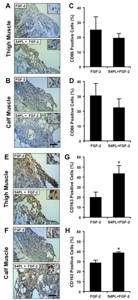
Figure 4. Syndesome and FGF-2 treated limbs have increased staining for M2 macrophage markers.
(A, B) Thigh and calf muscle sections immunostained for CD86 (M1 macrophage marker). (C, D) Quantification of percentage of CD86 positive cells in the thigh and calf muscle. (E, F) Immunostaining for CD163 (M2 macrophage marker) in the thigh and calf muscle. (G, H) Quantification of percentage of CD163 positive cells in the thigh and calf muscle sections. *Statistically different from FGF-2 only group (p <0.05; n = 8). Scale bars = 125 μm and insets are magnified by threefold.

