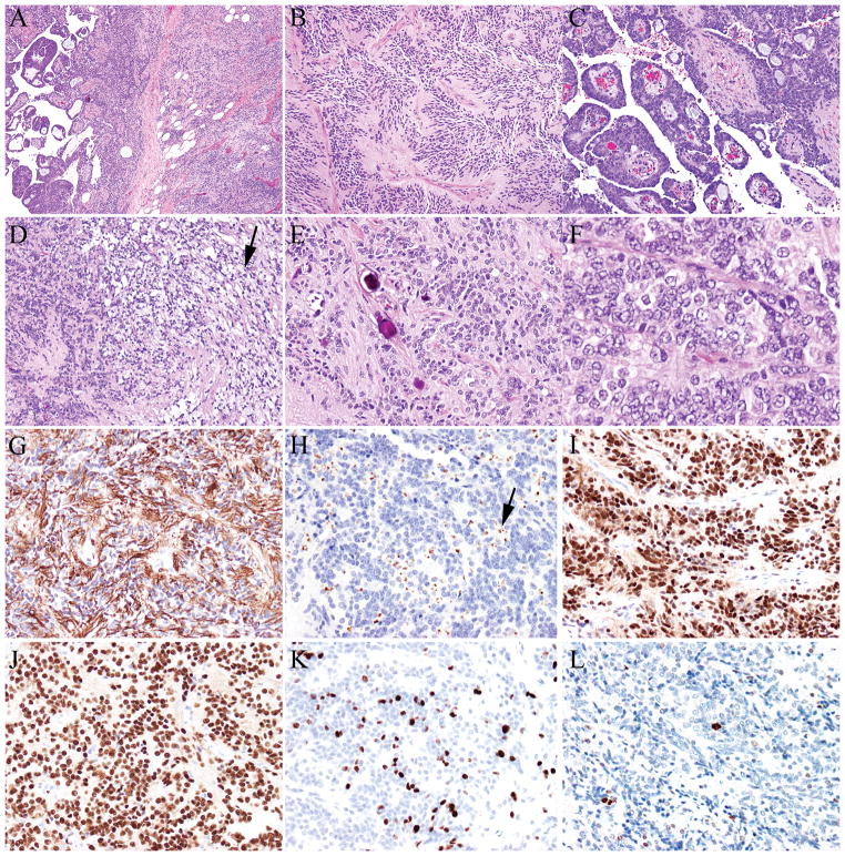
FIGURE 1.
A–F, Anaplastic ependymoma (A) showing multiple architectural patterns, including classical areas with perivascular pseudorosettes (B), papillary (C), and clear cell areas (D, black arrow). Psammoma bodies (E) were present. Some areas showed increased cellularity, nuclear atypia, and elevated mitotic activity (F). Tumor cells were positive for glial fibrillary acidic protein (G), epithelial membrane antigen (perinuclear dot-like pattern, black arrow) (H), estrogen receptor (I) and progesterone receptor (J), and demonstrated a high proliferative index (K- Ki-67/MIB-1). Phosphohistone H3 (pHH3) highlighted mitotic figures (L).

